Fetal Well Being Scan/Growth Scan
The growth scan, sometimes called the wellbeing scan or positioning scan, takes place when the mother is between 23 and 40 weeks pregnant. It checks how well the baby is growing and the position in the uterus (womb).
During the scan the doctor:
- Measures the baby's head, abdomen and thigh bone
- Assesses the amount of amniotic fluid around the baby
- Observes the baby's activity
- Measures the blood flow in the umbilical cord using doppler ultrasound
- Records the position of the placenta.
The measurements of the head, abdomen and leg allow the doctor to estimate fetal weight. All the measurements are plotted on a chart against the normal range and to assess the fetal growth. Because babies grow at different rates from week to week, a series of scans can be more helpful than just one.
Why is a growth scan required?
If the baby has an average head size, but a big abdomen, he/she may just be getting a good food supply from the placenta. If the mother has diabetes in pregnancy (gestational diabetes) she may have a big baby (macrosomia). This happens when the mother's blood sugar levels have risen too high. This can make the baby measure larger than expected for all parameters.
If the baby has an average head size and a small abdomen,, this may simply indicate a small, healthy baby. Occasionally, though, this can be a sign that the baby is not growing properly. The scan may also show that the amount of amniotic fluid is low. The two sometimes occur together.
If the baby is smaller than expected, he/she may have a low birth weight. To find out why the baby is small, the doctor might ask for a Doppler scan.
Scans are usually accurate for assessing the baby's size in the first half of the pregnancy. By the time the mother is in the later stages of pregnancy, scans continue to be accurate, as long as the baby is small or of average size. The closer it gets to the due date, and the bigger the baby is, the harder it will be to record measurements.
The baby's head may be too low in the pelvis in late pregnancy to get a measurement. Even if the baby's abdomen can be measured, it's very difficult to take other factors into account, such as how long the baby is.
Why is a scan even needed in the third trimester?
The most common reason for a scan in the third trimester is to check that the baby is growing normally. The mother will be offered a growth and fetal wellbeing scan between 28 weeks and 32 weeks of pregnancy. This will show the doctor how the baby is growing.
The mother will get another growth scan and colour Doppler studies closer to thedue date (if required), between 36 and 40 weeks to:
- Check the position of umbilical cord
- Measure the amount of amniotic fluid
- Check the placental position and maturity
- Know the baby's position and weight
- Check the baby's wellbeing and circulation
- Assess how a previous caesarean scar appears
The mother might get additional scans in thethird trimester if:
- The baby isn't moving as well or as often as he should be
- The baby is in the breech, oblique or transverse position
- The mother is carrying twins or more
- The amount of amniotic fluid is more or less than it should be
- The baby feels smaller or larger than expected for her gestational age
What can third trimester scans detect?
The third trimester scans will look at the following:
The baby's wellbeing
This is measured by his biophysical profile. A healthy baby:
- Stretches and flexes
- Moves his/her arms and legs frequently
- Opens and closes his/her hands
- Makes breathing movements
The doctor will observe the baby doing all these activities on the scan.
The doctor will observe the baby doing all these activities on the scan.
- Whether the dates are right: After 20 weeks, babies become more individual in size and shape. If the baby is smaller or bigger than average at, say 34 weeks, it doesn't mean the baby is younger or older. The due date needs to be established by 20 weeks.
- Where bleeding is coming from: Bleeding in the third trimester may be coming from the cervix or further inside the womb. A scan can reassure that the baby is not affected by the bleed but the scan can rarely see the cause of it. The scan can detect the cause only if it is due to a low-lying placenta.
- How much the baby weighs: The bigger the baby and the nearer to term, the harder it becomes to assess the weight.
What happens during a foetal growth scan?
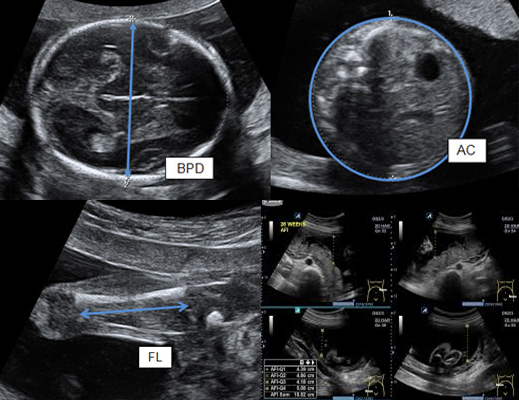
During the fetal growth scan, various measurements are taken of the fetus. The measurements are plotted on a growth chart, according to the number of weeks pregnant that the patient is at the time of the scan (gestational age).The main fetal measurements taken for a growth scan include:
- Biparietal diameter (BPD) measures across the head
- Head Circumference (HC) – measures around the head
- Abdominal Circumference (AC) – measures around the abdomen
- Femur Length (FL) – measures the length of the thigh bone
An estimate of fetal weight (EFW) can be calculated by combining the above measurements. The EFW is plotted on a graph to help determine whether the fetus is average, larger or smaller in size for its gestational age. If the fetal weight estimate is below the bottom 10 per cent line on the graph, it is considered to be small for gestational age (SGA). If the fetal weight is above the top 10 per cent line on the graph, it is considered to be large for gestational age (LGA).
It is important to note that repeated ultrasound measurements of the same fetus can vary and the estimated fetal weight may be incorrect by as much as 20 per cent.
A fetal growth scan does not routinely check the baby for abnormities. An ultrasound scan to look for major fetal abnormalities is routinely performed earlier in pregnancy between 18 and 20 weeks gestation
