Gandhinagar Fetal

Medicine Centre

Specialist In:
FETAL VIABILITY SCAN    |    FIRST TRIMESTER SCAN/NT SCAN    |    FETAL ANOMALY SCAN    |    FETAL ECHO    |    FETAL WELL BEING SCAN    |    3D/4D SCANS    |    INVASIVE TESTING    |    BLOOD TESTS    |    FETAL INTERVENTIONS

ABOUT DOCTOR

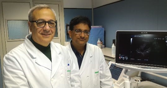
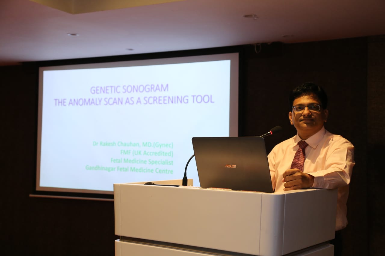
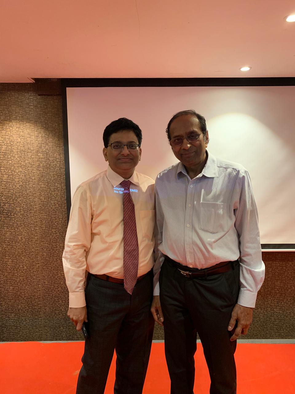
Dr Rakesh Chauhan Fetal Medicine Specialist, MBBS,M.D.(GYNEC),special trainings in field of Fetal medicine. He is accredited from Fetal Medicine Foundation, UK, Diploma in Invasive ultrasound from Italy. .

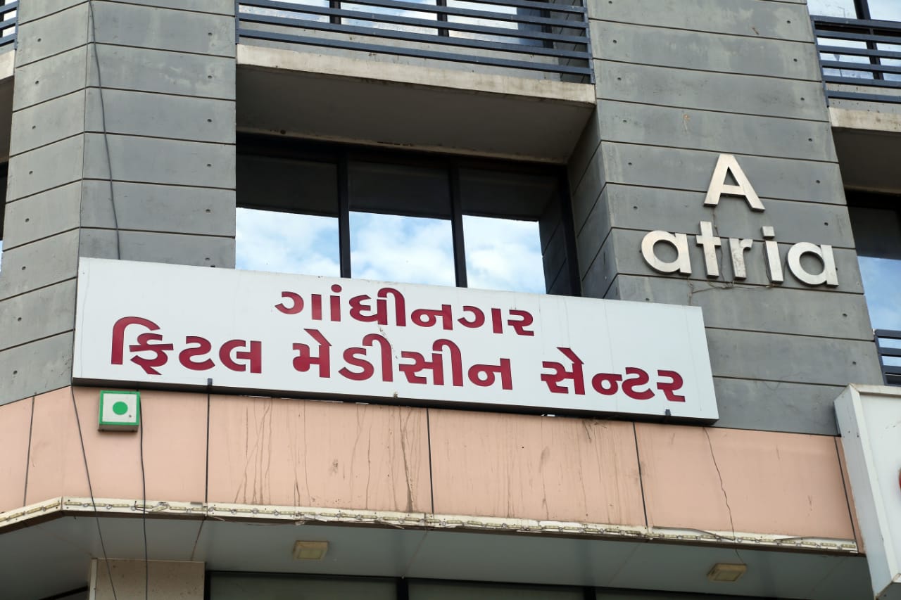
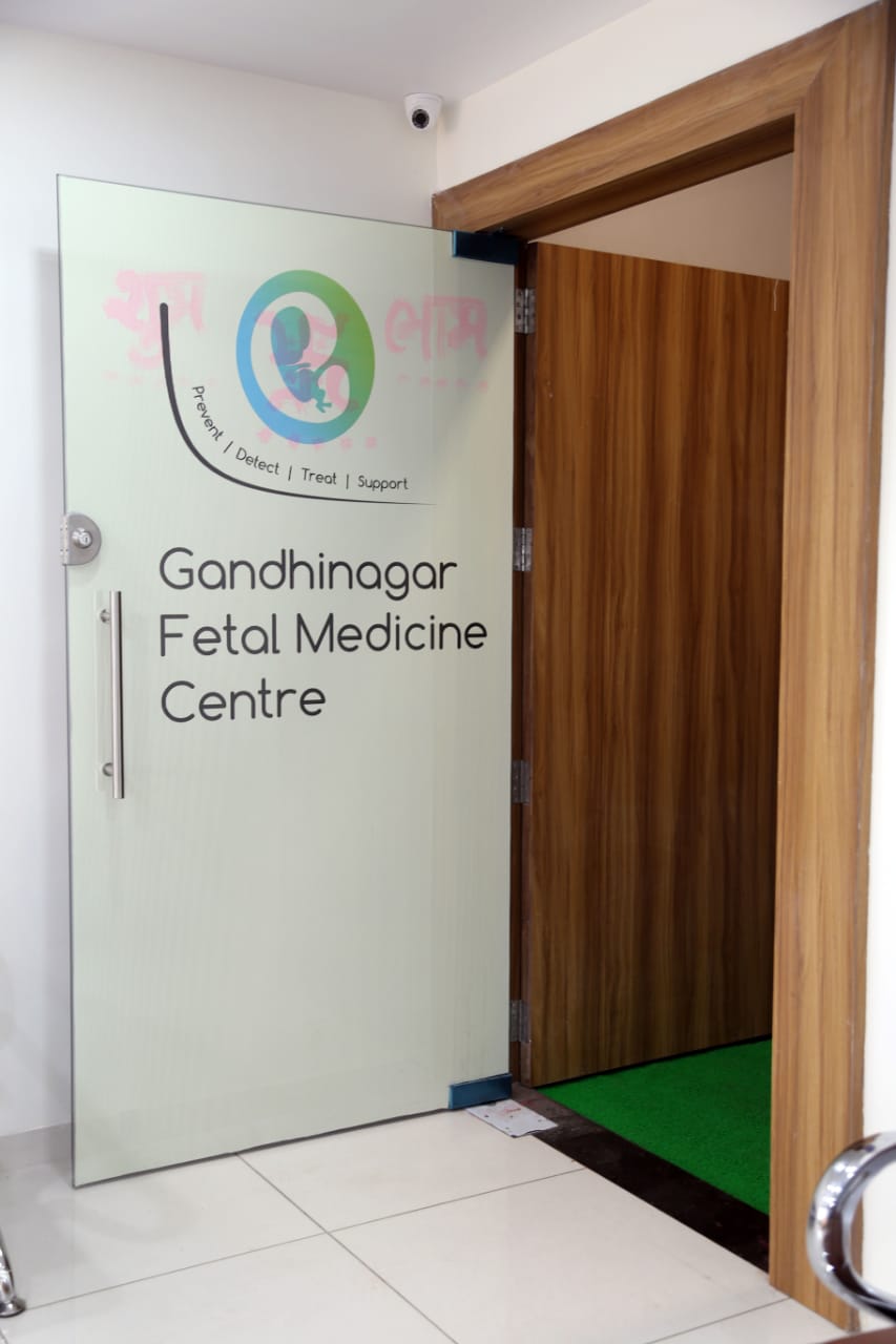
With objective of providing Fetal Medicine Service to the publics of Capital of Gujarat Gandhinagar,and surround districts,he has establish gandhinagar fetal medicine center

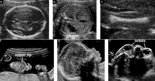
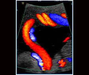
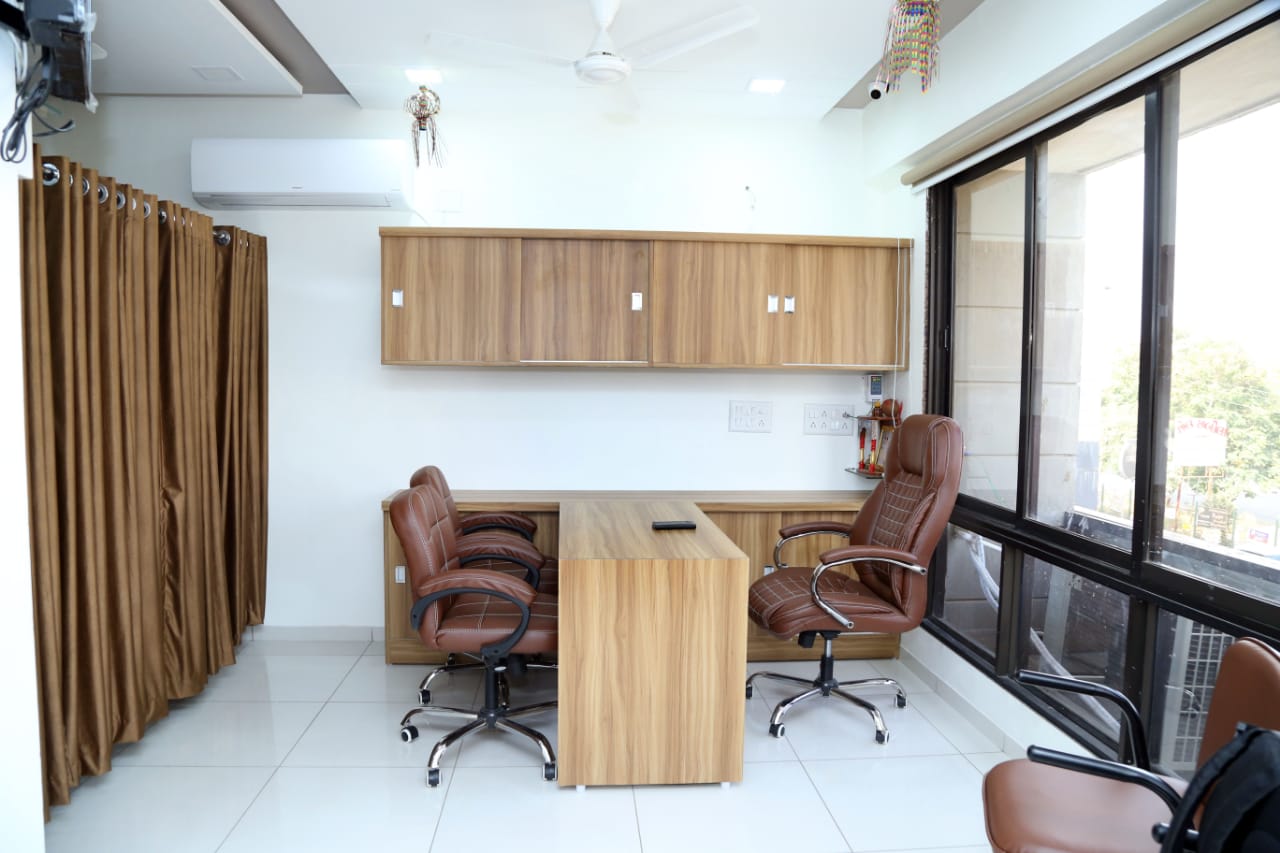
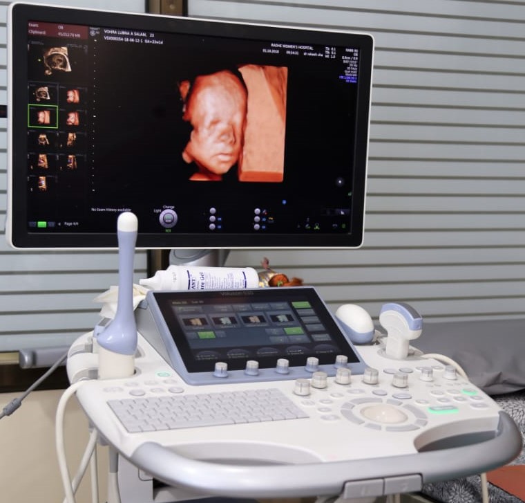
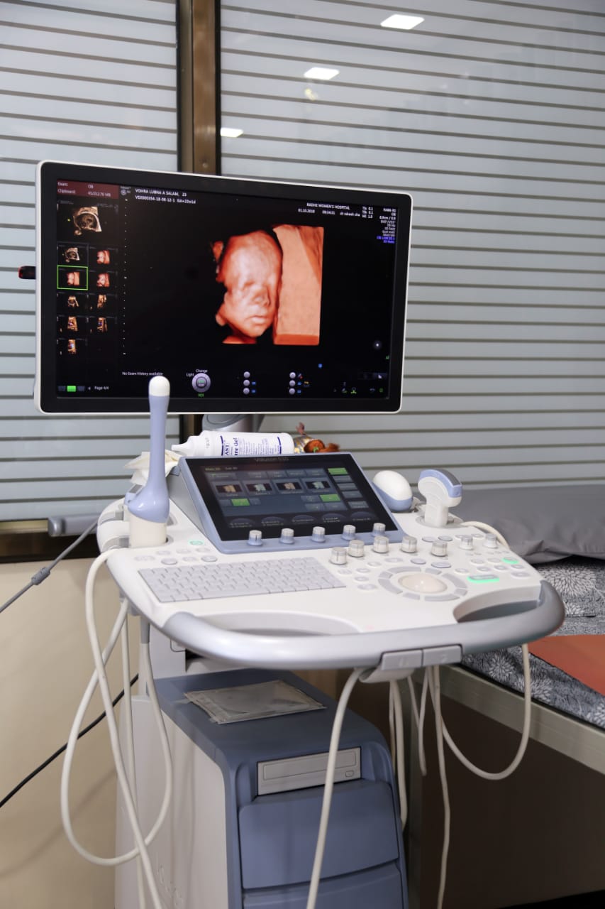
Gandhinagar Fetal Medicine Centre is first of its kind superspecialty centre in Gandhinagar dedicated to fetal care during pregnancy. Ultrasound scans are indispensable non-invasive tool which helps to assess fetal health.Understanding the unborn has been a challenge to a medical field. Fetal medicine is a highly specialized branch of obstetrics (pregnancy related) dealing with all matters that affect the health of fetus from conception to delivery.

It is specially deals care of fetus – identification and treatment of any problems which may arise to the fetus during pregnancy. Having a baby is one of the most joyous and exciting things in one’s life. The nine months of journey is very precious.

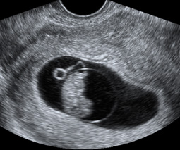
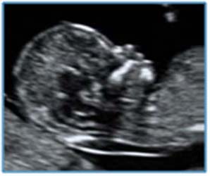
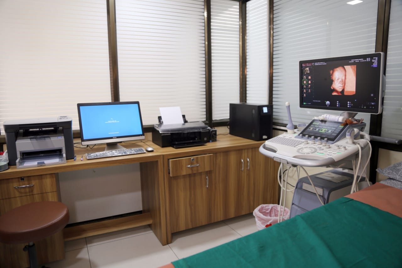
As a part of antenatal care all pregnant mother are offered ultrasound scans to monitor the baby’s health, development and check for any serious abnormalities. Early, more accurate screening gives peace of mind during pregnancy.

Ultrasound scanning uses sound waves therefore not harmful to mother or baby. As far as we know baby is unaware that scan is taking place. During pregnancy 3 scans are recommended. One during 3 month NT scan, one during 5th month anomaly scan and the third one around 8- 9 months "growth & fetal well-being scan".

Our Services

Which Services We Provide

department

Fetal viability Scan:

This scan is to confirm that pregnancy is inside the uterus,viability,number and also to look for adjacent organs. Sometimes it may be necessary to carry out TVS. This scan is especially useful for women who are experiencing bleeding or pain in the pregnancy and those who have had previous miscarriages or ectopic pregnancie

department

Fetal Trimester Scan/NT Scan:

The purpose of this scan is to: Demonstrate that the fetus is alive. Date the pregnancy from the measurement of the size of the fetus. Diagnose or exclude some major fetal abnormalities (upto 75% of anomalies can be excluded at this stage). Diagnose multiple pregnancies and determine the chorionicity (to decide whether identical or non-identical foetuses and this crucial for the management) Measure the nuchal translucency, to look for fetal nasal bone and in few high risk cases to study the blood flow through the heart valve and flow in ductus venosus .The scan done by a specialist who is certified from the fetal medicine foundation UK.These are used for calculating the chance that the fetus has Down’s syndrome and other major chromosomal abnormalities. Combining the ultrasound findings with measurement of two hormones produced by the placenta will achieve close to 95% detection rate (with 2.5% false positive rate) of Down’s syndrome. Basic details about pregnancy such as last menstrual period,mother’s age,weight,and medical/obstetric history also need to be included for the risk calculation.

department

Fetal Anomaly Scan:

This is a detailed examination that check for physical problems in the fetus. Fetal growth, amount of amniotic fluid and the position of the placenta will also be checked with this scan.It is best carried out around 20 weeks.Upto 95% of structural anomalies can be excluded from this scan. Basic study of Fetal heart is also done with anomaly scan and it is sufficient in all low risk pregnant women. However, in few situations detailed study of fetal heart is needed (known as Fetal ECHO). It is important to remember that scan does not pick up all problems in the baby. Measurement of the blood flow in the uterus is helpful to to determine risk of developing pre-eclampsia and small baby. Measurement of the length of the cervix will help to determine risk of premature birth.


department

Fetal Echo:

This is a detailed examination of fetal heart and this can be performed at the time of anomaly scan. Majority of the structural anomalies of the heart (upto 95%) can be detected from this study.The common indications are: Suspected fetal heart disease on scan. Previous child with heart problem. Family history of congenital heart disease. Pregnant mother with diabetes or on some medications(anti epileptic etc). Raised nuchal measurement or increased risk of chromosome problem. Other abnormalities detected during scan.


department

Fetal Well Being Scan/Growth Scan:

This scan is to study the growth of baby and also to study the blood flow (doppler)to test and well being of fetus to check whether the developing baby is healthy inside the woomb or not? . Therefore, this is scan is very useful in deciding timing of delivery especially in high risk cases (fetal growth restriction, problems in mother etc.) Frequent scans may be required for patients with special cases like when pregnant mother has high BP diabetes, kidney problems, multiple pregnancies, small baby, previous bad outcomes and so on.


department

3D/4D:

This scan shows 3 dimentional view of the baby and the best done around 28 weeks of pregnancy.3D is still colour picture and 4D is moving (live) images of the baby.With very high qualities of images,it is possible to clearly see babys mouth,lips,nose,eyes and facial expressions.Sometimes an excited family members claim that they can already see family resemblences!Baby may be sucking thumbs,yawning,smiling and this can be recorded on a DVD or still image pictures can be printed to ensure parents can cherrish thememories for years to come . In addition to enjoing the viewes of the baby,this scan is very usefull for the detailed assesment of few fetal anomalies(brain,face,spine ,limb problems etc.)

department

Invasive Testing:

The purpose of this scan is to: Demonstrate that the fetus is alive. Date the pregnancy from the measurement of the size of the fetus. Diagnose or exclude some major fetal abnormalities (upto 75% of anomalies can be excluded at this stage). Diagnose multiple pregnancies and determine the chorionicity (to decide whether identical or non-identical foetuses and this crucial for the management) Measure the nuchal translucency (fluid behind the fetal neck), to look for fetal nasal bone and in few high risk cases to study the blood flow through the heart valve (to look for tricuspid regurgitation) and flow in ductus venosus (in fetal liver).

The scan done by a specialist who is certified from the fetal medicine foundation UK.These are used for calculating the chance that the fetus has Down’s syndrome and other major chromosomal abnormalities. Combining the ultrasound findings with measurement of two hormones produced by the placenta (by doing blood test from the mother) will achieve close to 95% detection rate (with 2.5% false positive rate) of Down’s syndrome. Basic details about pregnancy such as last menstrual period,mother’s age,weight,and medical/obstetric history also need to be included for the risk calculation.


department

Blood Test:

Combined screening for chromosomal done between 11-14 weeks (detection rate of >90%) Triple test/Qudaruple test- done between 15-21 weeks (detection rate of 80%). NIPT (Non-invasive prenatal testing): Free fetal DNA can be extracted from maternal blood test.As early as by 11 weeks it is possible to diagnose Downs syndrome/other chromosomal anomalies with > 99% detection rate. In future it would be possible to diagnose genetic disorders as well. Parental Karyotyping, other genetic tests.

department

Fetal Interventions:

Various foetal procedures are performed such as, Selective fetal reduction:In cases of multiple pregnancies the fetal numbers can be reduced to desired number.This will substantially reduce the complications such as abortion, preterm delivery, cerebral palsy, financial burden etc. Fetal blood transfusions: Intrauterine foetal blood transfusions are very effective to treat foetal anaemia due to various causes. Fetal shunting:Sometimes shunting procedure needs to be done to treat fluid collection in lungs,bladder (vescico- amniotic shunt) etc.


Get in Touch

VIVO  Healthcare - Whats App